
Introduction
At Ephysioeeds Academy, our mission is to provide valuable knowledge and support for individuals and families facing the challenges of clubfoot. Clubfoot, medically known as Congenital Talipes Equinovarus (CTEV), is a complex foot deformity that, if left untreated, can hinder mobility and cause pain. Our goal is to shed light on clubfoot, its origins, causes, and the importance of early intervention.
Understanding Clubfoot
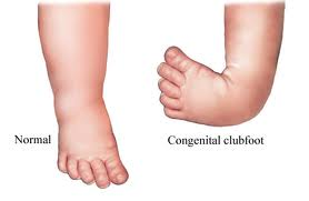
Clubfoot is a congenital deformity characterized by the misalignment of foot structures, involving bones and soft tissues in the hindfoot, midfoot, and forefoot. This condition presents as an inward turning of the midfoot (adductus and cavus) and a varus hindfoot, with the foot fixed in a downward-pointing (equinus) position at the subtalar joint. The affected foot is often shorter, and the calf circumference is smaller compared to a normal foot.
Key Facts About Clubfoot
- Clubfoot can affect one or both feet, with around 50% of cases being bilateral.
- It is more common in males than females, with a reported male-to-female ratio ranging from 1.6:1 to 3:1.
- While most cases of clubfoot are isolated, it can occasionally be associated with other congenital malformations or syndromes.
Causes of Clubfoot
The exact causes of clubfoot are not fully understood. Approximately 80% of cases are idiopathic (meaning the cause is unknown), while the remaining 20% are linked to other conditions, such as Spina Bifida, Cerebral Palsy, and Arthrogryposis. There appears to be a combination of genetic and environmental factors at play in clubfoot development.
One potential risk factor is maternal smoking during pregnancy, and there is also a genetic predisposition, particularly if one or both parents have had clubfoot.
Clinical Presentation and Examination
Recognizing clubfoot involves observing certain signs and symptoms:
- Smaller, stubby feet with a shortened first metatarsal ray.
- Equinus deformity (heel pointing downward) with inversion of the heel and varus of the forefoot.
- Concave and elevated medial border of the foot, with a convex and depressed lateral border.
- Prominent posterior heel tuberosity, which can be challenging to palpate.
- Callosities on the dorsal aspect of the fifth metatarsal.
Lack of Knowledge and Skilled Clinicians
In underdeveloped countries, many clinicians lack the qualifications and experience needed to treat clubfoot effectively. This results in misinformation and alternative treatments, including religious healings and surgery, being sought by parents. A lack of awareness contributes to children going untreated, perpetuating the misconception that clubfoot cannot be treated.
Conservative Treatment Options
- Ponseti Method: The widely accepted treatment involves manipulations by a skilled physiotherapist, serial castings, and a minor surgical procedure to release the Achilles tendon. When correctly applied and administered promptly, this method can lead to up to 90% recovery of normal foot alignment.
- French Method: An alternative technique, the French Functional (Physical Therapy) Method, is less common and involves daily manipulations, muscle stimulation, and foot immobilization using nonelastic adhesive strapping. Research indicates that results are slightly less favorable compared to the Ponseti Method due to the extensive parental involvement required.
Surgical Intervention
Surgery for clubfoot was more prevalent in the past but is now generally considered a secondary option. Early surgical approaches included soft tissue release techniques. However, long-term follow-up studies revealed complications such as stiffness and weakness in the foot and ankle. As a result, non-invasive methods are now preferred initially.
Idiopathic and Secondary Clubfoot
- Idiopathic clubfoot is the most common form, often with a hereditary component. It can range from untreated to successfully treated, with various degrees of impairment.
- Secondary clubfoot is associated with other diseases or conditions, typically neurological (e.g., Spina Bifida) or syndromic (e.g., Arthrogryposis).
Physiotherapy Management
Physical therapy plays a crucial role in clubfoot treatment:
- Bracing is essential for recovery, particularly for maintaining foot correction.
- Manual Therapeutic Techniques include soft tissue massage, stretching, and joint mobilization by a skilled therapist to improve foot alignment, mobility, and range of motion.
- Therapeutic Exercises focus on regaining range of motion and strengthening foot and lower extremity muscles.
- Stretching and Strengthening Exercises target specific muscle groups to address varus or valgus deformities.
- Electrical Stimulation (ES) is beneficial, particularly for acquired deformities.
- Taping Techniques can aid in maintaining foot alignment.
- Neuro-muscular Re-education (NMR) helps restore stability and improve movement techniques.
Conclusion
At our Academy, we believe in a compassionate approach to clubfoot treatment. Early diagnosis and intervention are crucial. We strive to bridge the knowledge gap and ensure that skilled clinicians are available to provide effective treatment. With the right care and support, individuals with clubfoot can lead fulfilling lives, emphasizing the importance of early, non-invasive intervention to achieve the best outcomes.
At Ephysioneeds Academy, we offer comprehensive training for physiotherapists looking to progress their skills. Our courses include dry needling certification, advanced physiotherapy techniques, and sports injury treatment. Our rehabilitation certification and physiotherapy training online provide flexibility and depth. We moreover specialize in manual therapy courses and soft tissue therapy certification. Explore our sports fellowship in physiotherapy and detailed A-Z knee and shoulder treatment courses. Additionally, the Tapedia Taping Encyclopedia is an essential resource for mastering taping techniques.